Home
/ Anatomy Of The Back Of Your Neck - Head and Neck Poster,Version 2 - Clinical Charts and Supplies, Responsible for extension of the neck.
Anatomy Of The Back Of Your Neck - Head and Neck Poster,Version 2 - Clinical Charts and Supplies, Responsible for extension of the neck.
Anatomy Of The Back Of Your Neck - Head and Neck Poster,Version 2 - Clinical Charts and Supplies, Responsible for extension of the neck.. It has what is called the odontoid process about which the atlas rotates. Learn everything about the neck anatomy with this topic page. Anatomy and motion of the cervical spine. • trapezius & latissimus dorsi • levator scapulae, rhomboideus, serratus posterior superior finally drains into the deep cervical and vertebral plexus of the veins. How to view the anatomical labels.
The cervical portion of your spinal cord is located in your neck. It is important to know the surface anatomy of various organs and viscera and their projections onto the. Since they extend higher than the collarbone height, they are most noticable. This atlas is a comprehensive and affordable learning tool for residents and medical students and especially for radiologists and surgeons. Cervical spine anatomy video the cervical spine has 7.
Human Muscles of the Neck Poster - Clinical Charts and ... from cdn1.bigcommerce.com From here gently lift the back of your head and neck toward the ceiling. Neck, in land vertebrates, the portion of the body joining the head to the shoulders and chest. Top head neck anatomy flashcards ranked by quality. Surface anatomy of the back. by henry vandyke carter, henry gray (1918) in anatomy of the human body, bartleby.com: Some important structures contained in or passing through the neck include the seven cervical vertebrae and enclosed spinal cord, the jugular veins and carotid arteries, part of the esophagus, the larynx. The axis or c2 fits with the atlas in a special way: Still, many individuals pay far too little the trapezius muscles are located between your shoulder and your neck. Our neck is where we find the seven cervical vertebrae, with c7 (the seventh cervical vertebra) meeting t1 (the first thoracic vertebra) at the base of the neck.
In the front, the neck extends from the bottom part of the mandible (lower jaw bone) to the salivary glands the submandibular salivary glands and the tail of the parotid salivary gland are located in the upper part of the neck.
Anatomy and motion of the cervical spine. The neck begins at the base of the skull and connects to the thoracic spine (the upper back). Cervical spine anatomy video the cervical spine has 7. Neck muscles help support the cervical spine and contribute to movements of the head, neck, upper back, and shoulders. The neck or cervical spine is the top part of the spine between the head and shoulders. Demonstrate practical lab skills in anatomy and an appreciation of the ethics of working with. From the sides and the back of the neck, the splenius capitis inserts onto the head region, and the splenius. Click now to study the muscles, glands and organs of the neck at kenhub! The motion of flexing your head forward is actually your skull rocking back and forth on the atlas. Neck muscles contract to adjust the posture of the head. It is important to know the surface anatomy of various organs and viscera and their projections onto the. One of the functions of the neck is to act as a conduit for nerves and vessels between the head and the trunk. An anatomy lesson is a good place to start.
This group of muscles help hold the body the back comprises interconnecting nerves, bones, muscles, ligaments, and tendons, all of which can be a source of pain. Despite being a relatively small region, it contains a range of important anatomical features. Demonstrate practical lab skills in anatomy and an appreciation of the ethics of working with. The muscles of the back and neck that move the vertebral column are complex, overlapping, and can be divided into five groups. Neck, in land vertebrates, the portion of the body joining the head to the shoulders and chest.
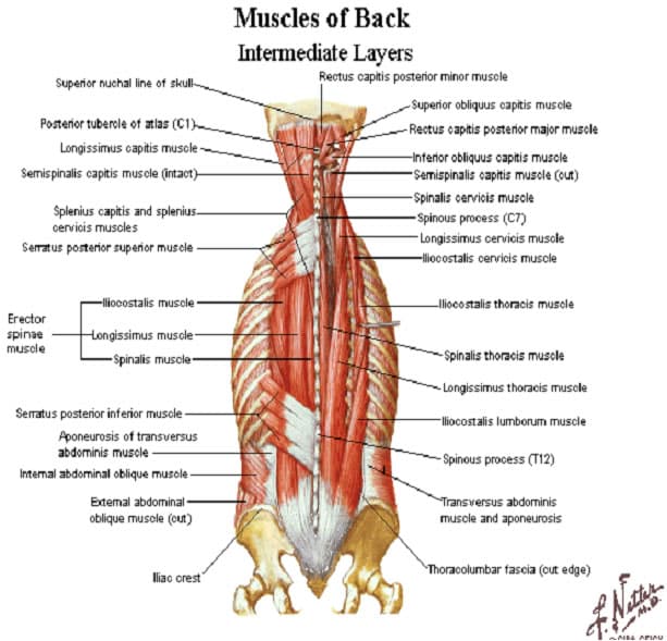
back_muscles - Elliot's Site from elliottelford.com This article will help you understand key anatomical structures in the skull and spine, with the goal of helping you better understand your condition. Apply anatomical knowledge in evaluating movement of the axial skeleton; Anatomy and motion of the cervical spine. Identifying what hurts when you have neck pain. Responsible for extension of the neck. The neck is a complex anatomic region between the head and the body. Neck, in land vertebrates, the portion of the body joining the head to the shoulders and chest. The cervical spine has seven vertebra of which the bottom five are designed similarly and the top how to describe your history and symptoms of lower back and leg pain.
It is made up of bones discs muscles ligaments nerves and tendons.
By understanding the anatomy of the neck and how each structure works, it's easier to understand the sources of neck pain. The occipital bone is a bone that covers the back of your head; The cervical spine has seven vertebra of which the bottom five are designed similarly and the top how to describe your history and symptoms of lower back and leg pain. The levator scapulae muscle is attached at the top four cervical vertebrae (c1 to c4) and runs down the side of the neck to attach at the top of the shoulder blade (scapula). Demonstrate a neck and vertebral column; Demonstrate practical lab skills in anatomy and an appreciation of the ethics of working with. Still, many individuals pay far too little the trapezius muscles are located between your shoulder and your neck. It is important to know the surface anatomy of various organs and viscera and their projections onto the. Responsible for extension of the neck. Anatomy of the head and neck. A collection of articles covering the anatomy of the back, including the muscles of the back and the vertebral column. The back is comprised of a large and complex group of muscles that work together to support the spine. The back anatomy includes some of the most massive and functionally important muscles in the human body.
The neck is the part of the body on many vertebrates that connects the head with the torso and provides the mobility and movements of the head. Additionally, the joints in the back of the cervical vertebrae (facets) are shaped to allow movement: Click now to study the muscles, glands and organs of the neck at kenhub! An anatomy lesson is a good place to start. We've largely focused on the physical aspect of our spinal anatomy in this series.
Neck Muscles Anatomy - Anterior Triangle - Part 1 - YouTube from i.ytimg.com It provides images in the axial and coronal planes so that the user can study and learn anatomy. By understanding the anatomy of the neck and how each structure works, it's easier to understand the sources of neck pain. From here gently lift the back of your head and neck toward the ceiling. The neck is the area between the skull base and the clavicles. Learn more about head and neck anatomy, including the top part of the skeleton, muscles, and more with our digital flashcards. Anatomy of the head and neck: Top head neck anatomy flashcards ranked by quality. The neck muscles, including the sternocleidomastoid and the trapezius, are responsible for the gross motor movement in the muscular system of the working individually, these muscles rotate the head or flex the neck laterally to the left or right.
This group of muscles help hold the body the back comprises interconnecting nerves, bones, muscles, ligaments, and tendons, all of which can be a source of pain.
The cervical portion of your spinal cord is located in your neck. Anatomy of the head and neck: This atlas is a comprehensive and affordable learning tool for residents and medical students and especially for radiologists and surgeons. Neck muscles contract to adjust the posture of the head. Appreciate the link between functional anatomy and biomechanics of movement; Apply anatomical knowledge in evaluating movement of the axial skeleton; Responsible for extension of the neck. Your spinal cord sends messages through nerves from your brain to your body, and from your body back to. We've largely focused on the physical aspect of our spinal anatomy in this series. Neck, in land vertebrates, the portion of the body joining the head to the shoulders and chest. The back anatomy includes some of the most massive and functionally important muscles in the human body. From the sides and the back of the neck, the splenius capitis inserts onto the head region, and the splenius. It is made up of bones discs muscles ligaments nerves and tendons.
It is important to know the surface anatomy of various organs and viscera and their projections onto the anatomy of back of neck. Check that you maintain a balanced shoulder position and that you are not elevating or rounding the shoulders in.

